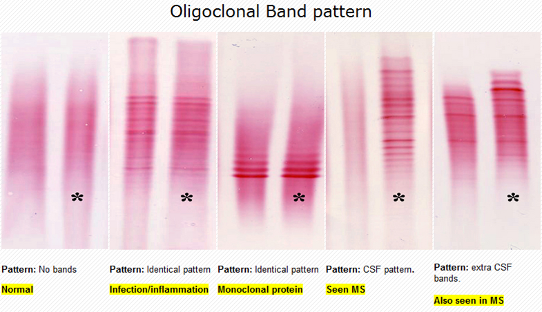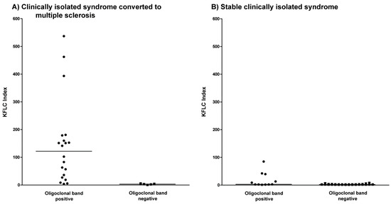

Oligoclonal bands are found in a variety of inflammatory and infectious CNS disorders, including paraneoplastic disorders, CNS lupus, neurosarcoidosis, and Behçet’s disease however, the majority of cases in which oligoclonal bands are seen are infectious. 36 While oligoclonal bands are detected in the majority of MS patients, the presence of oligoclonal bands is not specific to MS.

CSF and serum samples are compared although there are multiple patterns of CSF oligoclonal bands, a significant pattern is one in which two or more bands are present in the CSF but absent in the serum. Immunoblotting is then used to detect separated IgG molecules. Oligoclonal bands are detected by isoelectric focusing methods that separate proteins in the CSF and serum, including IgG. IgG index is obtained by calculating the ratio between CSF and serum IgG after correcting for albumin concentrations in the CSF and serum. The presence of oligoclonal bands and elevated IgG index in the CSF have long been used to aid in the diagnosis of MS. Oligoclonal bands and IgG elevation in the cerebrospinal fluid (CSF) provide further evidence to support a viral etiology in MS. Steven Jacobson, in Human Herpesviruses HHV-6A, HHV-6B & HHV-7 (Third Edition), 2014 Oligoclonal Bands and IgG Index in MS A band of red Hb can also be seen in the β region before staining of the electrophoretic gel.īridgette Jeanne Billioux. Confirmation that this band is Hb depends on visual examination of the CSF for blood or hemolysis (red color). This band of Hb should not be confused with clonal immunoglobulin in CSF. Presumably, it represents clonal proliferation of plasma cells in the body with passive movement of the monoclonal antibody from blood into CSF.Īnother distortion of the electrophoretic pattern occurs with release of hemoglobin (Hb) into the CSF, resulting in a major band in the β region close to transferrin ( Fig. However, this finding is not indicative of intrathecal synthesis and, thus, is not a positive result for oligoclonal bands. The oligoclonal bands in Figure 47.9A are discrete and clearly separate from one another.Įxamination of serum and CSF sometimes shows a clonal band of immunoglobulin present in both ( Fig. After all these landmark proteins of CSF are recognized, oligoclonal bands should be evaluated in the γ region, which normally has only a small quantity of polyclonal immunoglobulins. C3 is also identifiable in CSF, along with a band of asialotransferrin in the β2 region.
OLIGO BANDS CSF POSITIVE AND OLIGO BANDS SERUM 2 PLUS
The higher molecular weight proteins in serum (α2 plus β-lipoprotein) are absent from CSF. The other significant band in CSF is transferrin because of its small molecular size that permits ultrafiltration from blood into CSF. Albumin is the predominant band in both fluids. CSF has a relatively higher concentration of prealbumin than does serum.

The CSF was concentrated, and the serum was diluted to achieve comparable amounts of stainable protein in each application. The protein electrophoretic pattern of CSF and serum from a patient with multiple sclerosis is shown in Figure 47.9A. Another possibility is an immunologic response to central nervous system infection thus, the interpretation of oligoclonal bands in CSF must be made in the context of other diagnostic findings, such as cell counts, viral serologies, bacterial cultures, syphilis testing, nucleic acid amplification tests for viruses, and so forth.

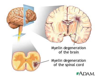
Oligoclonal bands as a marker of synthesis of immunoglobulins in the central nervous system suggest the possibility of an autoimmune process, such as a demyelinating disease. The interpretation of CSF protein electrophoresis requires simultaneous electrophoresis of the patient’s serum to ensure that findings in the CSF are unique and do not reflect just the passive transfer of clonal immunoglobulins from the blood. McPherson MD, MSc, in Henry's Clinical Diagnosis and Management by Laboratory Methods, 2022 Oligoclonal Immunoglobulin Bands in Cerebrospinal FluidĮvaluation of intrathecal synthesis of immunoglobulin is aided by high-resolution electrophoresis of cerebrospinal fluid (CSF) protein that spreads out the γ region to display individual bands of different immunoglobulin clones.


 0 kommentar(er)
